
The patient and his story
The 69-year-old patient, immunocompromised and suffering from cancer, presented to the outpatient department of the dermatological clinic in Ludwigshafen due to a “ ulceration of the left forearm which progressed rapidly over a few days », report dermatologists.
Minor trauma, probably an insect bite, had preceded the operation.
Two weeks of outpatient treatment with amoxicillin did not improve the situation. The man reportedly had type 2 diabetes and hypertension; he also had an aortic valve implanted transfemorally. His medications: metformin, acetylsalicylic acid, a statin, antihypertensives and hydroxycarbamide.
The results
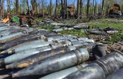
- Ulceration approximately 10 × 8 cm with livid wound margin, ambient redness and central necrosis on the left forearm.
- Palpation of lymph nodes unremarkable, as well as peripheral blood circulation, motor skills and sensitivity.
- Microbiological results: detection of Staphylococcus aureus, Enterobacter ludwigii And Pseudomonas aeruginosa.
- Absence of hepatitis B or C infection.
- Elispot (Enzyme Linked ImmunoSpot) tuberculosis not interpretable in the laboratory due to CMML.
- Chest x-ray and ultrasound of the abdomen and lymph nodes were unremarkable.
- Mycological diagnosis (native preparation and fungal culture) negative.
- Histological result “in principle compatible” with the diagnosis of suspected pyoderma gangrenosum (PG).
Treatment, evolution and diagnosis
- Oral treatment with 80 mg of prednisolone and topical treatment with disinfectant compresses and an ointment containing clobetasol propionate.
- Due to the rapid progression of the size of the ulceration, recommended treatment was initiated with adalimumab and immunoglobulins.
- Simultaneous meropenem and linezolid with increase in CRP (CRP 124 mg/dl).
- Rapid evolution despite therapeutic escalation.
- Thromboembolectomy due to acute decrease in perfusion due to thrombosis of forearm vessels.
- Histology of the embolus of the left radial artery: angio-invasive mycosis; PCR detection of Rhizopus arrhizus And Aspergillus niger.
- A new treatment of the spindle biopsy taken initially from the edge of the ulcer: a few isolated mycelia (PAS and Grocott staining); Aspergillus not detectable histologically.
- Forearm amputation due to rapidly progressive decrease in perfusion and formation of necrosis.
Despite intensive therapeutic measures, lung and liver damage could not be avoided. The patient refused further diagnosis to detect the pulmonary pathogen and ultimately died due to multiorgan failure.
Diagnosis: primary cutaneous mucormycosis with hematogenous dissemination in the context of an underlying immunodeficiency linked to CMML, type 2 diabetes, without forgetting iatrogenic immunosuppression
Discussion
According to dermatologists, mucormycosis is a rare and potentially fatal angioinvasive infection caused by fungi in the order Mucoralesgenre Rhizopus being the most common pathogen. Although rarer than invasive candidiasis or invasive aspergillosis, mucormycosis is still the second most common mold infection in Europe, explains Dr Patrick Schwarz (IV Medical Clinic of Hematology and Oncology, University Hospital of Giessen) in a recent article on mucormycosis published in a journal. In European countries, patients at risk are particularly immunocompromised patients, mainly those with underlying hematological diseases. In patients with HIV or AIDS, mucormycosis is rather rare. In addition to hematological disorders, type 2 diabetes is the second most common disease in patients with mucormycosis in Europe.
The most common form is rhinocerebral mucormycosis, but pulmonary, primary cutaneous or gastrointestinal forms are also possible, explain Drs Stefanie Lischker and Christiane Gieding. Hyphal invasion into the vascular system results in hematogenous dissemination.
While secondary cutaneous mucormycosis usually results from a rhinocerebral or pulmonary form, the rare primary cutaneous mucormycosis is caused by direct inoculation of spores, for example during minor trauma, burns or iatrogenically through injections.
According to Stefanie Lischker and Christiane Gieding, primary cutaneous mucormycosis usually occurs in acute or chronic leukemia and presents in a non-specific form, very similar to PG. The extremities are most often affected.
Detection by histology, fungal culture and PCR is often difficult. Mushroom cultures could be falsely negative in up to 50% of cases. Histology with PAS and Grocott staining plays an important role, especially for primary cutaneous mucormycosis.
Treatment of primary cutaneous mucormycosis includes, in addition to surgical debridement, elimination of risk factors and rapid administration of systemic antifungals. The lethality of invasive mucormycosis, however, would be greater than 50%, even if treatment is started early.
This case study was originally published on Univadis.de.


