(AFP) – It’s nuclear but it’s medicine: advances in imaging, already crucial for detecting and treating breast cancer, now extend to “tracers” of tumors and metastases in the ‘body.
pixelfit / Getty Images
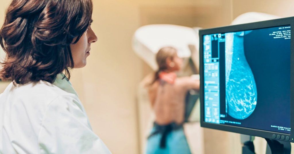
The principle: weakly radioactive molecules, called radiotracers, are injected intravenously and, once in the blood, spread throughout the patients’ bodies.
“Part of the molecule emits radiation, which allows us to have images; another attaches to a receiver,” summarized Romain-David Seban, nuclear doctor and researcher at the Institut Curie, during a press conference ahead of Pink October.
Behind a “concept that is sometimes a little scary because there is the word + nuclear +” hides a technique for better treatment, explained this expert before the annual breast cancer awareness campaign.
With more than 61,000 new cases each year in France, breast cancer remains the most common among women, and the deadliest, with around 12,000 deaths per year.
Depending on their stage of development, their location in the organ, the cells from which they spread or the presence of hormonal receptors, breast cancers differ. Therapeutic responses too.
To refine the diagnosis, detect possible metastases or measure or even predict the response to treatments, maximum information is needed, and nuclear medicine can play a role.
This imaging already used is not invasive, because it does not require a biopsy, and provides information on the entire body, where a biopsy is very targeted, its specialists praise.
Dr Seban cited the example of a patient “with triple negative breast cancer, who came for a follow-up assessment to see if her disease was circumscribed or not”. “As there were no metastases, she was eligible for treatment with surgery, chemotherapy and radiotherapy, plus immunotherapy as her tumor was relatively aggressive,” he explained.
Technological progress is reducing the time it takes to obtain images after the injection of a radiotracer, ensuring less waiting and less fatigue for patients, observe the specialists.
For the moment, nuclear doctors mainly use a machine called a PET scanner, with a radiotracer targeting the cells of the body that consume the most sugar – which include cancer cells.
– “Revolution” –
But there are sometimes false positives, or, on the contrary, certain metastases not visualized.
Other avenues are therefore being tested for the future, in the hope of better diagnoses, or even predictions of reactions to treatments.
The Institut Curie is banking in particular on a tracer that attaches to specific cells in the tumor microenvironment, fibroblasts.
Under study in clinical trials for triple negative breast cancers, the most aggressive, a new generation radiotracer (the “FAPI”) could better identify metastases, evaluate the effectiveness of a treatment or detect a relapse early, according to its specialists.
Researchers are also testing a tracer targeting hormone receptors, particularly estradiol, to find out if they are present in the breasts of certain patients because they determine the effectiveness of hormone therapy.
“More and more trials are seeking, by combining imaging and nuclear medicine, to see which patients will respond very well to treatment or for whom we could reduce chemotherapy and/or immunotherapy tomorrow, or even who could no longer respond. operate. It’s a revolution,” said Anne Vincent-Salomon, director of the Women’s Cancer Institute, co-created by Curie, Paris Sciences et Lettres (PSL) and Inserm.
In the future, a weapon used against other cancers (thyroid, neuroendocrine tumors or prostate) could also be used against breast cancer.
The mechanism is reminiscent of ballistics: a radiotracer specific to the tumor microenvironment is coupled to molecules capable of destroying these diseased cells. This “vectorized internal radiotherapy” is being studied for breast cancer.
“Image is revolutionizing cancer care. Far from a photograph that describes, we are in the image that treats, even predicts,” underlined Steven Le Gouill, director of the Curie hospital complex.
In addition to its increasing analytical finesse, it provides faster responses to caregivers – capabilities that artificial intelligence should multiply.
Relax