Scientists at the University of East Anglia have developed advanced MRI technology to diagnose a common heart problem faster and precisely than ever.
Aortic stenosis is a progressive and potentially fatal condition, affecting around 300,000 people in the United Kingdom. It affects about five percent of the 65s in the United States, with an increasing prevalence in advanced age.
A new study, published today, reveals how a four -dimensional flow MRI (4D flow) can diagnose more reliable aortic stenosis than current ultrasound techniques.
The higher accuracy of the new test means that doctors can better predict when patients need surgery.
We hope that the breakthrough could help save thousands of lives in the United Kingdom only.
The main researcher, Dr. Pankaj Garg, of the Norwich School of the UEA and a consultant cardiologist at the Norfolk and Norwich University Hospital, said: “Aortic stenosis is a common but dangerous heart disease.
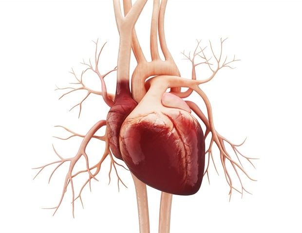
“This happens when the aortic valve, the main valve of leaving the heart, stiffens and shrinks. This causes a reduction in the blood flow of the heart in the rest of the body.
“Symptoms include chest pain, a rapid floating heart rate and a feeling of vertigo, shortness of breath and fatigue – especially with activity.
“For the moment, doctors use an ultrasound to diagnose the condition, but this can sometimes underestimate the severity of the disease, delaying vital treatment.
“The 4D flow MRI is an advanced cardiac imaging method that allows us to look at the blood flow in three directions over time – the fourth dimension.
-“We wanted to see if it could provide a more precise and reliable diagnosis than a traditional ultrasound. »»
The team examined 30 patients diagnosed with aortic stenosis using both traditional ultrasound analyzes (echocardiography) and advanced IRM imaging.
By comparing the results, they evaluated the method identified with more precision patients who need a timely valve intervention in a timely manner.
They validated their results by comparing them to real clinical results over a period of eight months.
The team noted that the 4D flow MRI technology offered more precise and reliable measures of blood flow through patients’ cardiac valves, compared to traditional echocardiography.
We hope that this breakthrough will transform the way cardiologists assess patients with aortic stenosis-leading to more timely interventions, unless complications and potentially thousands of lives saved in the United Kingdom. “”
Dr Pankaj Garg, the UEA Norwich Medicine School
This research was led by the EUA in collaboration with the Norfolk and Norwich University Hospital NHS Foundation Trust, the University of Sheffield, the San Juan Hospital in Dios (Spain), the University of Chieti-Pescara (Italy), the University of Leeds and Leide University Medical Center (The Netherlands).
He was funded by Wellcoma.
“The four -dimensional flow provides an incremental diagnostic value on echocardiography in aortic stenosis” is published in the journal Open heart.


