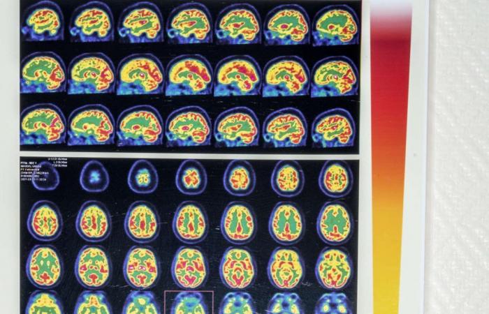
Functional neurological disorders (FND) are one of the main reasons for consultation in neurology. Loss of use of a limb, speech disorders, abnormal movements or even dissociative functional crises, their manifestations are varied. While imaging shows no damage to the central nervous system to explain them, these symptoms are very real. Until now, scientists have struggled to understand and explain their mechanisms. Béatrice Garcin, professor of neurology at the Avicenne hospital in Bobigny, takes stock.
In the absence of brain lesions, what does imaging say about TNF?
Data varies depending on the methodology used. But functional MRI studies show hyperactivation of regions processing emotions, notably fear, at the level of the amygdala – strongly connected to motor regions of the brain – even when these patients are subjected to a positive emotion. And this, especially since there was trauma during childhood. While the brain matures until the age of 25-30, this suggests that the brain of a child who grew up in insecurity has developed in survival mode to face danger, like in an animal. in nature: it overreacts to the slightest stimulus.
In functional abnormal movements, patients have movements that resemble voluntary movements, but do not have the perception that they are the ones who made the movement. They are said to have a loss of sense of agency. This sense relies heavily on a region called the “right temporoparietal junction”, and which integrates both the control of movement and the perception of the movement performed. In these patients, several studies have shown that this region dysfunctions, notably with hypoactivity. This clearly proves that the person does not “produce” their symptom, while many caregivers still doubt it – especially since we do not find these abnormalities on imaging in patients who malinger. Swiss researchers are evaluating the usefulness of using transcranial magnetic stimulation to stimulate this right temporoparietal junction, in the hope of promoting better agency.
From there, can we deduce operating mechanisms?
Functional imaging gives us clues to better understand TNF, but, for the moment, we do not fully understand the links between the hyperconnectivity of certain areas and the triggering of symptoms. We have identified risk factors (abuse, psychological disorders, head trauma, neurological diseases) and factors precipitating the appearance of TNF (new psychotrauma, intense stress, physical shocks), but we do not know how to explain their entanglement with functioning. of the brain. However, we know that precipitating factors determine the manifestation of the pathology. An injury, for example, can trigger a motor TNF such as loss of mobility of a limb or abnormal movement.
You have 44.99% of this article left to read. The rest is reserved for subscribers.


