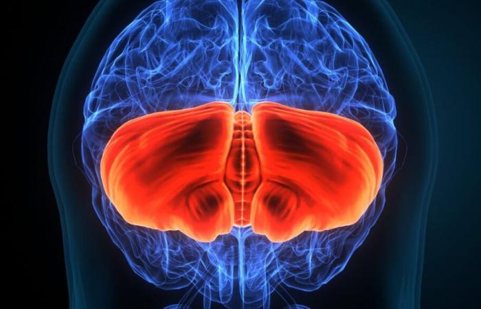
The placebo effect describes a phenomenon in which the simple anticipation of a beneficial effect produces this very effect. It is both the oldest and one of the most effective medical interventions. And, despite this, very little known until now. However, in an article recently published in the journal NatureChong Chen, a neuroscience researcher at the University of North Carolina, and his colleagues describe a simple method to produce a placebo effect that relieves pain in mice, allowing them to identify the neural circuit involved. The population of neurons in question is located in the limbic system – which is not in itself a surprise, because this region of the brain is known to be involved in particular in the processing of pain – but these neurons also communicate with parts of the brainstem and cerebellum. And this is unexpected, because these regions are usually associated with more basic functions, such as the coordination of movements…
The reality of the placebo effect has long been debated, in particular because it is difficult to know whether an improvement in symptoms is due to or whether it is due to a favorable evolution in the patient’s condition, such as that. happens in many cases. But today it is no longer contested. And particularly in one area, that of pain relief. In this matter, the placebo effect comes down to this simple fact: believing that the pain will recede effectively results in analgesia, which we call “placebo analgesia”. We know today that this phenomenon notably involves natural analogues of morphine that our body produces, enkephalins or beta-endorphin (thus, when we administer to animals a molecule – naloxone – which prevents the action of these molecular messengers, the placebo effect disappears).
How to study the placebo effect in the laboratory?
In 2021, researchers wanted to know which regions of the brain were responsible for the placebo effect. To do this, they brought together all the data obtained to date thanks to brain imaging, which highlighted two major facts: first, the areas involved in the perception of pain see their activity decrease during placebo effect. But, more surprisingly, this is also the case of the cerebellum, which is usually rather involved in the coordination of movements… How can we explain this?
Imaging studies, useful as they are, do not offer as fine a resolution as modern methods that can trace neural circuits in mice, and such techniques had not previously been applied to the study placebo analgesia. We obviously cannot teach mice to wait for events to occur through verbal instructions, but it is possible to create anticipation in them through conditioning, that is, learning by association. The protocol developed by Chong Chen and his colleagues is based on a device composed of two compartments: the floor of the first is heated to 30°C, the second to 48°C. After a few days, the mice which entered the room at 48°C therefore expected to find a floor with painful contact, while those entering the compartment at 30°C expected to encounter a painless floor.
Then, on the day of testing, the mice are placed in the two-compartment apparatus. Everything is identical to previous days, except for one detail: this time, the floors of the two compartments are heated to 48°C. However, although the stimulus was identical for all the mice, those who learned to associate one of the compartments with the pleasant temperature of 30°C this time preferred to spend time in it, despite the temperature at now much higher. These mice apparently suffer less, lick their paws less, do not jump in the air as much or rear up as much as in the other compartment. As if expecting not to suffer actually diminishes their perception, and this effect persists for several days, while also applying to other types of painful stimuli than heat.
© Cerveau et Psycho, according to Jeffrey S. Mogil, Placebo effect involves unexpected brain regions, Nature, 2024
The neurons of “analgesic anticipation”
The neuroscientists then sought to identify areas of the brain that were active when the mice expected to suffer less. And they limited their research to brain areas connected to a particular region called “rostral anterior cingulate cortex”, or CCAr. Indeed, previous data had shown that the latter, an integral part of the limbic system, played a role in placebo analgesia. Three brain structures were thus identified: the striatum, the thalamic and subthalamic nucleus and a pair of structures in the brainstem called “pontal nuclei” (nP).
However, the nPs form the connection between the cerebellum and the cerebral cortex. What is surprising is that they are not considered to belong to the classic pain treatment network. Experts responsible for evaluating the article by Chong Chen and his colleagues before publication noted that these nuclei connect to the cerebellum, and therefore recommended that we examine the activity of the neurons most representative of the latter, Purkinje cells, in mice anticipating placebo analgesia. The scientists then identified a group of Purkinje cells that activate when the animal expects this relief. The activity of this group of cells depended on the circuit connecting the rostral anterior cingulate cortex to the nP…
This is not the first animal model of placebo analgesia, nor the first use of conditioning to study this phenomenon. But Chong Chen and his team provide here the most careful explanation yet, mobilizing essentially all the modern high-resolution techniques currently available to study neuronal circuits in mice (calcium imaging to analyze activity neuronal activity in awake mice, electrophysiology to record the activity of neurons in brain slices, optogenetics to activate or block neurons on command using light pulses, or even RNA sequencing of cells individuals to observe gene expression).
Have we identified the exact neural circuits underlying this effect in humans? We will still have to wait before concluding. And keep in mind that the underlying mechanisms of this phenomenon are probably more complex in humans since they involve anticipations formed according to verbal instructions and social influences, beyond simple conditioning.
What is particularly interesting in this study is the demonstration that, among all the potential brain areas, it is the pons nuclei and the cerebellum which are responsible for the anticipation of placebo analgesia, a phenomenon which we would have more readily attributed to so-called “higher” structures of the central nervous system responsible for coordinating complex cognitive functions. It will now be a matter of interpreting this discovery, which could lead to a re-evaluation of the neuroanatomy of pain.
It is the pons nuclei – a basic structure of the brain – and the cerebellum that are responsible for anticipation of placebo analgesia. While we would have thought more of so-called “higher” structures responsible for coordinating complex cognitive functions…


