⇧ [VIDÉO] You might also like this partner content
The first comprehensive mapping of the vinegar fly’s neurons and synaptic connections was recently completed. This monumental feat is the work of the Flywire Consortium, an international collaboration. This connectome constitutes a major advance in the field of neuroscience and will undoubtedly allow a better understanding of the human brain.
Treatments for neurodegenerative diseases such as dementia (including Alzheimer’s) or Parkinson’s disease remain somewhat experimental and lack effectiveness. The complexity of the human brain and the lack of understanding of the circuits that make it up make the development of targeted treatments difficult. “ To understand how the brain generates perceptions, thoughts, and actions, it is crucial to grasp how information travels from sensory receptors to motor outputs “, says Dr. Mala Murthy, co-leader of the Flywire project at Princeton University.
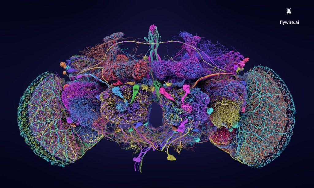
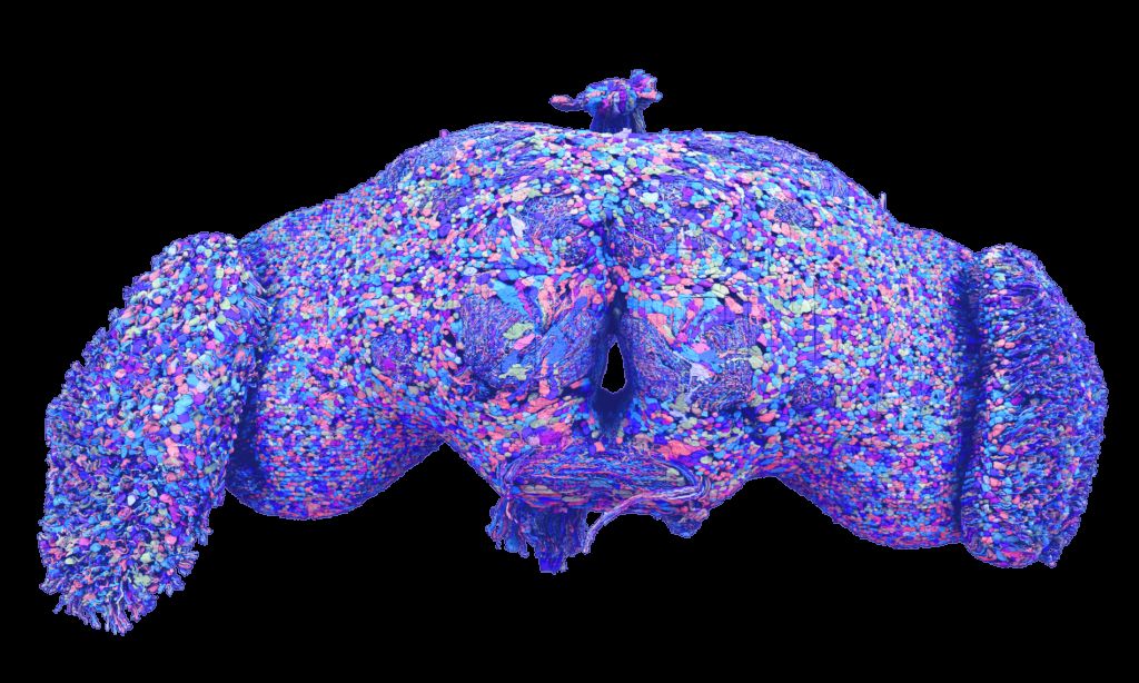
Previous research had partially mapped the brain of a fruit fly larva, comprising 3,016 neurons, as well as that of a nematode worm, with 302 neurons. A partial map of the brain of a mouse had also been published. However, this new complete connectome of a vinegar fly (or Drosophila), produced as part of the Flywire project, is the first to detail the entire adult brain of the insect. Scientists have identified the position, shape and connections of the 139,255 neurons in the brain of the Drosophila melanogasteras well as the 50 million synapses that connect them.
This feat required ten years of work for researchers from the Flywire Consortium, from the Medical Research Council (MRC) molecular biology laboratory, Princeton University, the University of Vermont and the University of Cambridge. The choice fell on adult Drosophila because of its importance in neuroscience. This fly, capable of walking and flying, also demonstrates complex behaviors such as learning and good memory. Males can even “sing” to attract females.
« Why be interested in the brain of a vinegar fly? “, asks Sebastian Seung, professor of computer science and neuroscience at Princeton University and co-leader of the Flywire project, in a press release. “ If we can understand one brain, it will necessarily teach us something about all brains. ».
A careful and complex process
To establish the connectome, the researchers began cutting the brain of a fruit fly, no larger than a poppy seed, into 7,000 sections. Each section was photographed using a high-resolution microscope, capturing structures with a minimum thickness of 40 nanometers. The resulting images were then assembled to form a three-dimensional representation of the brain.
Analyzing nearly 100 terabytes of image data to trace the path of 139,255 neurons and their 50 million synaptic connections constitutes a complex process. The researchers therefore resorted to artificial intelligence. Gregory Jefferis, from the Cambridge Molecular Biology Laboratory, explains: “ We started by manually identifying neurons, but that would have required more than 4,000 years of human work ».
However, the AI models, although trained based on the scientists’ work, made errors in reconstructing the neurons, requiring manual intervention to correct them. The team then opened the data to the scientific community, allowing researchers and volunteers to collaborate (since 2019) to reread and annotate the results of the AI models.
Once the connectome was completed, the scientists were able to label the different types of neurons and synaptic connections, classifying more than 8,400 different cell types. “
This dataset is like Google Maps, but for a brain: the wiring diagram between neurons reveals which structures in satellite images of Earth correspond to streets and buildings », explains Dr Philipp Schlegel from the MRC laboratory. This new map is made available to the global scientific community for any future research.
See also

Researchers have classified more than 8,400 different cell types. © Tyler Sloan for FlyWire, Princeton University
The brain diagram produced also allowed researchers to make promising discoveries. For example, they identified neurons which combine different types of information and others, called diffusers, which coordinate the activity of neuronal circuits. The neurons involved in movement have been located at the base of the brain, while those processing vision are located laterally. The use of AI has also led to a better understanding of the role of neurotransmitters in the amplification or inhibition of neuronal messages. Details of the Flywire project have been published in nine articles in the journal Nature.

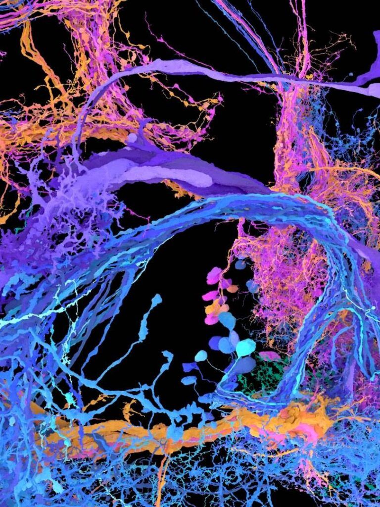
Image showing the brain cells of the vinegar fly’s auditory circuits, as mapped and annotated by FlyWire. © Princeton University
According to the researchers, the Drosophila connectome will serve as a springboard for mapping more complex brains, notably the human brain, with its 86 billion neurons and one hundred thousand billion synapses.
To date, only one cubic millimeter of the human brain has been mapped. The next step for the Flywire Consortium is full mapping of the mouse brain, an ongoing project expected to be completed in five to ten years. “ The task remains titanic to decipher how the brain functions », concludes John Ngai, director of the Brain Initiative at the United States National Institutes of Health.