Scientists have developed a rapid and non -invasive blood test that can detect Parkinson’s disease before the tremors start. By measuring RNA fragments that reflect the pathology of the brain, the test offers new hope for early diagnosis and targeted intervention.
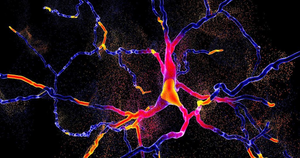
Study: Blood test of Parkinson PRERESYMPYMATIME quantifying repetitive sequence reasons in transfer RNA fragments. Image credit: Kateryna Kon / Shutterstock
In a recent study published in the journal Aging of natureThe researchers have evaluated whether a blood test measuring the fragments of nuclear and mitochondrial transfer RNA (ARNT) could precisely detect Parkinson Presmpomatic (PD) disease.
Background
What if we could detect the PD before the start of a single tremor? MP is the second most common neurodegenerative disorder in the world, affecting more than 10 million people worldwide depending on the largely cited estimates and causing progressive movement and cognitive deficiencies. Current diagnostic methods are often reactive, identifying the disease after significant brain damage has already occurred. Invasive tests and incoherent biomarkers more emphasize early diagnosis. Transfer RNA fragments (TRF), small non -coding RNA parts generated by enzymatic cleavage, emerge as potential indicators of neurological disorders. Their levels move in response to mitochondrial dysfunction and neural stress, the two characteristics of the PD. However, additional research is necessary to validate their diagnostic power.
About the study
Researchers have carried out a multi-cohort analysis using a small RNA sequencing and a quantitative polymerase chain reaction (QPCR) to explore the specific TRF diagnostic potential in the PD. They analyzed the samples of cerebrospinal fluid, blood and brain of patients with PD, Alzheimer’s disease and healthy witnesses, including post-mortem samples of the Brain Bank of the Netherlands (NBB) and living donors of the initiative of Parkinson’s progression markers (PPMI). The study was focused on two TRF families: RGTTCRA-TRFS with nuclear nuclear, derived from the transfer RNA and marked by a specific repetitive motif ((A / G) GTTC (A / G) A), and Mitochondriaux (MT-TRF), from mitochondrial genomes. A relationship between the two was calculated to normalize the differences between individuals.
Using the PPMI data set, they evaluated this report in patients at an early stage, to transport mutations and prodrome. Using the double QPCR, they also validated the results of fresh blood samples (Shaare Zedek medical center cohort) and post-mortem brain tissues (Nih NeurobiBank). In addition, they used an automatic learning model (GBM) on a gradient scale to compare the predictive precision of the TRF ratio with traditional clinical scores such as the unified scale of Parkinson disease (UPDRS) and the Hoehn and Yahr scale (H&Y). Additional experiences included overexpression and knockout of angiogenin (Ang), an arnt flashing enzyme and ribosomal profiling to examine the biological effects of the accumulation of RGTTCRA-TRF on protein synthesis.
Study results
The study revealed separate changes in transfer RNA fragments associated with MP. In the cerebrospinal fluid, patients had high levels of RGTTCRA-TRFs and a decrease in MT-TRF levels compared to witnesses and people with Alzheimer’s disease. This unique TRF profile was consistent in both sexes and showed no overlap with the signatures of Alzheimer’s disease. Likewise, the brain tissues of the substantia nigra (the most affected region in the MP) have shown high levels of RGTTCRA-TRF correlated with the presence of Lewy bodies, protein aggregates which are a brand brand of the disease.
Blood analysis supported these results. In the post-mortem samples, the RGTTCRA-TRF were significantly high while the MTOTs have been reduced. Patients in early stage and transporting mutations had a higher RGTTCRA / MTTR ratio than healthy carriers of the same mutation. This model was coherent in ethnic circles, although slightly less distinct among black participants, parallel to the trends in their clinical scores. Above all, the GBM model using the TRF ratio has reached diagnostic accuracy (area under the curve (AUC)) of 0.86, outperforming traditional clinical scores (AUC 0.73). The TRF signature has also distinguished prodromal patients, those with early and non -motor symptoms, healthy witnesses.
Double QPCR tests have confirmed that this report could reliably separate patients from witnesses both in fresh blood and post-mortem brain samples. In addition, RGTTCRA-TRF levels decreased after the deep treatment of brain stimulation (DBS), aligning the relief of clinical symptoms. Patients treated with DBS have shown a reduction in RGTTCRA-TRF and a decrease in angiogenin expression (Ang), indicating that the DBS can remove the production of TRF or modify their regulation.
Biological analyzes have provided an overview of the potential pathogenic role of RGTTCRA-TRFS. These fragments have shown a strong complementarity of sequence to the ribosomal RNA and to a fragment derived from the essential arnt leucine for the translation of proteins (leucag3′-TRF). Interaction modeling has suggested that RGTTCRA-TRFS could be binded to both, creating a “double locking” mechanism which alters the initiation and elongation of translation. The ribosomal profiling of depolarized neuroblastoma cells revealed a reduced association of RGTTCRA-TRF with ribosomes, supporting their role in the disturbance of translation. Förster resonance energy transfer imaging (freight) has confirmed proximity to proximity between RGTTCRA-TRFS and ribosomes in living cells.
Conclusions
To summarize, this study presents convincing evidence that the transfer of RNA fragments (in particular, RGTTCRA-TRFS and MT-TRFS) can be used as non-invasive non-invasive blood biomarkers for the PD. Their separate diagram allows a precise diagnosis even at the prodromal stages, surpassing traditional clinical notation. The Dual QPCR test is rapid, profitable and sensitive, which makes it very applicable in clinical environment. In addition, these TRFs can play a role in the progression of the disease by interfering with protein synthesis. Although the results require validation in larger and more diverse cohorts, in particular in under-represented ethnic groups, this biomarker strategy offers a promising route to previous detection, better surveillance and more effective therapeutic intervention in MP.


