Gilbert Bernier
Credit: ResearchGate
A team led by researcher Gilbert Bernier, from the University of Montreal and the Maisonneuve-Rosemont Hospital Research Center, has developed a method for performing retinal transplants using stem cells on blind minipigs. These animals showed signs of restored vision, which is a promising development for humans as well. The results of this study were published on December 5, 2024 in the journal Development.
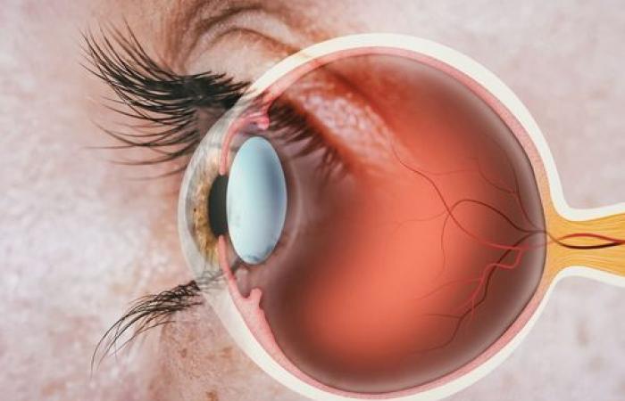
Millions of people around the world suffer from degenerative retinal diseases. In most cases, vision loss is due to damage to the macula, a central region of the retina rich in cone photoreceptors – cells essential for perceiving colors and fine details. Although quality of life is greatly impaired, there is currently no approved treatment to replace the damaged macula.
To achieve this breakthrough, Professor Bernier’s team designed a method to induce stem cells to form cell layers reproducing the structure of the human retina. These stem cells, called human induced pluripotent stem cells, are “reprogrammed” adult cells capable of differentiating into any cell type. Using these cells, the team created “retinal sheets” enriched with immature versions of cone photoreceptors, which can become mature when grown in the laboratory.
After creating these leaflets in the laboratory, the scientists undertook the delicate task of grafting them onto minipigs with damaged macula.
The professor explains that, “to get as close as possible to a human clinical application, we chose minipigs, whose eye size is close to that of humans and whose weight is similar. Thus, all the surgeries in our study could be performed by a retinal surgeon.
New connections between neurons and transplanted cells
Once the leaflets were transplanted, the research team found that the retinal grafts were able to integrate with the damaged tissue in the minipigs’ retinas. Encouragingly, the latter showed signs of visual recovery: new neuronal connections formed between the grafted photoreceptor cells and the minipigs’ neurons. Additionally, photoreceptor neuronal activity was detected in the grafted area when the animals were exposed to bright light.
Faced with the urgency of developing treatments for vision loss, researchers around the world are exploring different approaches to repair a damaged macula.
“Teams use dissociated photoreceptor cells; others create microdissected retinal organoids, mini-organs grown in the laboratory, says Gilbert Bernier. On the other hand, our method allows the spontaneous formation of flat retinal tissue, already polarized and organized, as in a human embryonic retina.” This method also makes it possible to produce large quantities of retinal tissue for transplants.
However, one of the limitations of this method is the difficulty in controlling the placement and orientation of the grafts during surgery. The macula is only four millimeters in diameter, about the length of a grain of rice.
“Properly orienting, placing and stabilizing the graft in the retina remains a major surgical challenge,” says the professor.
His team is now working to improve the success rate of transplants, in particular by developing an experimental retinal surgery device to guarantee adequate orientation and implantation of the graft at the precise location of the retinal disease.
Although many challenges remain, this study highlights the potential of retinal leaflet transplantation to treat degenerative retinal diseases.
Health


