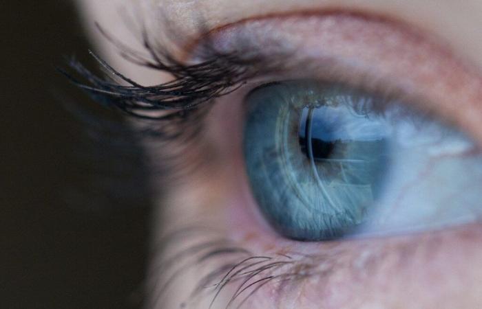
Three people suffering from severe visual impairment » and having received a stem cell transplant saw their eyesight improve “ considerably » and has been doing so for over a year. In a fourth person, this improvement was only temporary. These four patients are the first to receive reprogrammed stem cell transplants to treat “ corneas[1] damaged ».
The researchers published their results in The Lancet [2]. According to Kapil Bharti, researcher at US National Eye Institutein Bethesda, Maryland, they are “ impressive ».
Reprogrammed cells
The outer layer of the cornea is maintained by a “ reservoir » of stem cells housed in the limbal ring, the dark ring around the iris. When this source is depleted, it causes a disorder called limbal stem cell deficiency (LSCD). Scar tissue covers the cornea, which can lead to blindness. This deficiency can result from ocular trauma or autoimmune or genetic diseases.
Treatments for DCSL are limited. They usually involve the transplantation of corneal cells derived from stem cells taken from a person’s healthy eye,” an invasive procedure with uncertain results “. When both eyes are affected, a corneal transplant from a deceased donor may be considered, with a risk of rejection.
Professor Kohji Nishida, an ophthalmologist at Osaka University in Japan, and his colleagues used another source of cells, induced pluripotent stem (iPS) cells, to perform the corneal transplants. To do this, they took blood cells from a healthy donor and “ reprogrammed to the embryonic state ”, before differentiating them in order to obtain “ a thin transparent sheet of corneal epithelial cells ».
A well-tolerated treatment
Between June 2019 and November 2020, the team recruited four patients: two women and two men aged 39 to 72, with DCSL in both eyes. After removing the layer of scar tissue covering their damaged corneas, the researchers implanted the “ leaves » of donor-derived tissue and placed a protective contact lens on top. Only one eye was operated on in each patient.
All four receivers showed a “ immediate improvement » of their vision and a “ reduction of the affected cornea area “. Improvements persisted in all but one recipient, who showed “ slight setbacks » during the one-year observation period.
Additionally, two years after the transplants, none of the recipients experienced serious side effects. The grafts did not generate tumors and did not show signs of attack by the recipients’ immune systems, even in the two patients who did not receive immunosuppressive treatment.
Upcoming clinical trials
For Kapil Bharti, “ it is not clear what caused the vision improvements “. It is possible that the transplanted cells themselves proliferated in the recipients’ corneas. But the vision gains could also be due to the removal of scar tissue before transplantation, or because the transplantation triggered the migration of the recipient’s own cells from other areas of the eye and rejuvenation of the cornea .
Professor Nishida says he plans to launch clinical trials in March to assess the effectiveness of the treatment. Several other trials based on iPS cells are underway around the world to treat eye diseases (see Adrenal glands, eyes, bone marrow… iPS cells, a Swiss army knife?). This year, these cells also made it possible to treat diabetes in a Chinese patient (see iPS: a young diabetic woman produces her own insulin).
[1] transparent outer surface of the eye
[2] Soma, T. et al. Induced pluripotent stem-cell-derived corneal epithelium for transplant surgery: a single-arm, open-label, first-in-human interventional study in Japan, Lancet https://doi.org/10.1016/S0140-6736(24)01764-1 (2024).
Sources : Nature, Smriti Mallapaty (08/11/2024) ; Wired, Mariam Khan (11/11/2024)


