
By the evening edition.
The Nikon Small World Competition, a competition which rewards the best photos taken with ultra-magnifying equipment, has released its 2024 prize list. The opportunity to nourish science and marvel at what the human eye cannot see. ordinary not perceive.
It is a world that is inaccessible to us. To the naked eye at least. But by using microscopes, small organisms are revealed, sometimes even showing a certain graphic grace. For fifty years, the optics and photography specialist Nikon has rewarded the most beautiful microphotographs, that is to say taken with magnifying equipment.
Here are the images that caught the eye of the jury made up of scientists this year. They combine poetry and real scientific interest. A list published this October 17, 2024.
Read also: Hippopotamus, deer, monkey… Who will be the winner of the funniest animal photos of the year?
Better understand Alzheimer’s
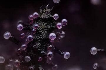
Last year, the competition highlighted a close-up of the head of a rodent’s optic nerve. This time, the first prize is awarded to Dr Bruno Cisterna, with the help of Dr Eric Vitriol, from Augusta University (United States). Their image, described as “revolutionary” by the organizers, shows cells differentiated from mouse brain tumors.
“This image reveals how disruptions to the cellular cytoskeleton – the structure and ‘highways’ called microtubules – can lead to diseases like Alzheimer’s and ALS [la maladie de Charcot] »develops the jury.
“One of the main problems with neurodegenerative diseases is that we do not fully understand their causes, says Dr. Cisterna. To develop effective treatments, we must first understand the basics. Our research is crucial to discovering them. And finally find a cure. The differentiated cells could be used to study how mutations or toxic proteins that cause Alzheimer’s disease change neuronal morphology. » This research could make it possible to develop and test treatments aimed at protecting neurons or restoring their function.
An electric arc
In second place, it is Dr Marcel Clemens, based in Italy, who is rewarded, in a very different field. His photo shows an electric arc “between a pin and a wire, produced by the application of a potential difference of 10,000 volts”.
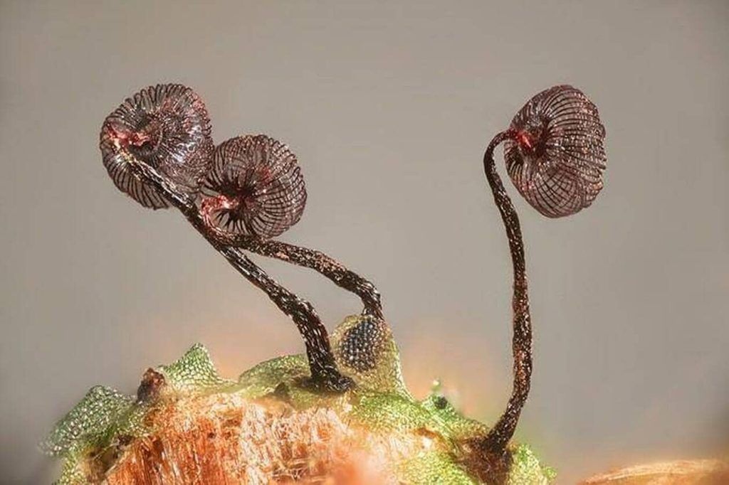
Zoom on a cannabis leaf
American Chris Romaine completes the podium with his ultra-zoomed image of a cannabis leaf. “The bulbous structures are trichomes or hairs and the bubbles inside are cannabinoid vesicles”specifies the organization.